

Posted by Sarah Collins
on September 12, 2026

Early, non-cavitated tooth decay can often be arrested or reversed through remineralization. Once the enamel surface breaks down into a true hole, the lost tooth structure cannot grow back and needs dental treatment.
So, is tooth decay reversible, or is that just wishful thinking? The honest answer is both yes and no, depending on what has happened beneath and on the surface of the enamel. A faint white spot may represent mineral loss while the outer enamel remains intact. A visible pit or hole is different because the tooth's structure has already been lost.
That distinction matters more than most online advice suggests. Remineralization can support early enamel lesions, but it can't rebuild a missing section of tooth at home. This guide explains how decay develops, where the reversible window ends, which fluoride and fluoride-free options have the strongest evidence, and what practical steps can help you protect enamel without fear or hype.
Introduction Is Tooth Decay Reversible
Many people hope a cavity will heal if they brush more carefully, stop eating sugar, or use a remineralizing toothpaste. That hope makes sense because the body can repair some early mineral damage. The confusion starts when the word cavity describes every stage of decay, from an invisible change inside enamel to a fully formed hole.
Early decay is a process, not an instant event. Acids can pull minerals from enamel, then saliva and topical ingredients can help return minerals while the surface remains intact. Once bacteria and acids create a break in that surface, home care may help protect the surrounding tooth, but it can't recreate the missing enamel.
The practical dividing line: mineral loss may be reversible before cavitation. A hole is structural damage, not just a mineral imbalance.
A white-spot lesion is often the classic early sign. It may look chalky, especially when the tooth is dry, because mineral loss changes how light moves through enamel. Yet a white spot isn't automatically decay. Developmental enamel changes and other conditions can look similar, so a dental professional should assess a new, changing, or uncertain mark.
The encouraging shift in modern dentistry is that early lesions can be managed rather than automatically drilled. Historical clinical evidence helped establish that reversals could be measured in real patients, not only in laboratory models, while current remineralization science clarifies why the process works. The aim isn't to promise a cure. It's to identify changes early enough to support the enamel that remains.
What Tooth Decay Really Is and How Enamel Loses Minerals
Think of enamel as a tightly packed brick wall. The bricks are mineral crystals, while the spaces between them influence how strong and smooth the surface feels. When acids repeatedly remove minerals, the wall develops microscopic gaps. If the environment improves, saliva can help refill some of those spaces.
Plaque bacteria use fermentable carbohydrates and produce acids. During an acid episode, the balance shifts toward demineralization, meaning minerals leave the enamel. Saliva helps buffer those acids and supplies calcium and phosphate, two materials enamel needs for mineral redeposition. Fluoride can also influence the mineral exchange by helping enamel resist acid and encouraging mineral return.
Why pH changes the direction
The mouth constantly moves between mineral loss and mineral gain. Enamel demineralization becomes more favorable below about pH 5.5, while returning above that critical point supports a shift toward remineralization, according to a review of the caries process and enamel repair (the pH threshold for enamel remineralization).
The brick-wall analogy helps here. Acid lowers the “repair conditions,” making it easier for minerals to leave. As saliva raises the pH and brings calcium and phosphate back into contact with enamel, the conditions become more favorable for redepositing mineral.
What makes the balance harder
Frequent acidic or sugary exposures give the mouth repeated opportunities to move toward demineralization. Reduced saliva can also make buffering less effective. Brushing removes plaque, but the larger goal is to reduce the conditions that let acid remain active around enamel.
For a broader look at the process, see this explanation of what causes tooth enamel loss. People who want to understand how oral conditions may relate to their routine can also explore an oral health test UK resource, while remembering that testing doesn't replace a dental examination.
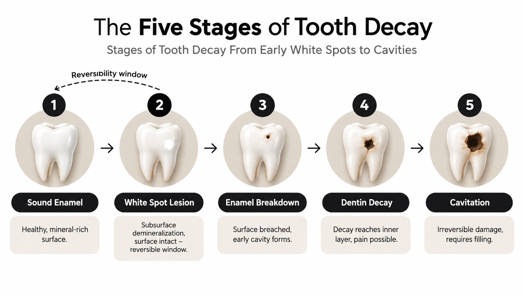
Stages of Tooth Decay From Early White Spots to Cavities
Decay usually develops along a progression, although the appearance and speed can vary. The most important question isn't whether a mark is white or dark. It's whether the enamel surface is still intact.
Stage one, sound enamel
Healthy enamel has a smooth, mineral-rich surface. It still experiences daily acid exposure, but saliva, cleaning, and mineral exchange can keep the overall balance stable. At this point, there's no decay lesion to reverse. The task is to preserve the surface.
Stage two, the white-spot lesion
An early white spot can reflect subsurface demineralization beneath an intact outer layer. Educational dental material describes these lesions as potentially involving about 30% to 50% mineral loss while remaining non-cavitated (development of the carious lesion and white-spot stage). Because the surface hasn't broken, minerals can still move back into the affected area.
A white spot may be easier to notice after drying, and it may not hurt. That absence of pain doesn't prove the area is harmless, nor does the spot alone confirm decay. A dentist may evaluate its location, texture, history, and appearance to distinguish early caries from developmental enamel changes.
Stage three, enamel breakdown
The reversibility window narrows sharply when the surface develops a pit, rough area, or hole. At that point, the enamel wall has been breached. Mineral support may still protect nearby enamel, but it can't replace the missing structure.
Stage four and beyond
If decay progresses inward, it can reach dentin, the softer layer beneath enamel. Sensitivity or pain may appear, but symptoms vary, so pain isn't a reliable staging tool. Deeper disease can involve the pulp and surrounding tissues, which requires professional assessment and treatment.
| Surface finding | What it may indicate | Can home remineralization restore lost structure? |
|---|---|---|
| Smooth, sound enamel | No visible lesion | Not applicable |
| Chalky white area with intact surface | Early, non-cavitated mineral loss | It may be arrested or reversed |
| Pit, rough break, or hole | Cavitated enamel | No |
| Deeper decay or persistent symptoms | Progression into inner tooth layers | No, professional care is needed |
The table is a guide to the structural boundary, not a way to diagnose yourself. A mirror can show a mark, but it can't reliably show whether the surface is intact.
When Tooth Decay Is Reversible and When It Is Not
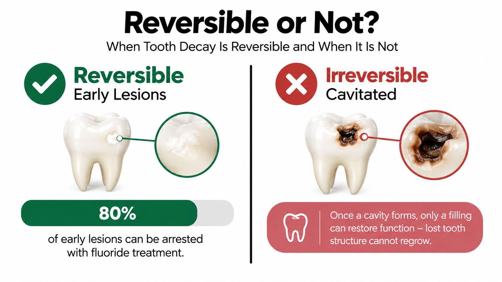
The clearest answer is simple: early, non-cavitated lesions may be arrested or reversed; cavitated decay is not reversible without dental treatment. Remineralization works with the enamel that remains. It doesn't regenerate a missing piece after the surface collapses.
Evidence supports realistic optimism rather than guaranteed results. A 2016 meta-analysis of professionally applied 5% sodium fluoride varnish found an overall remineralized-enamel-caries rate of 63.6%, with a 95% confidence interval of 36.0% to 91.2%, in the included studies (the meta-analysis of professionally applied fluoride varnish). This figure describes study results for early lesions under treatment conditions. It isn't a promise that every white spot will reverse.
Earlier clinical evidence also matters. In a 3-year randomized study published in 1998, people using sodium fluoride experienced statistically more caries reversals than those using placebo, particularly among participants who already had at least one lesion at baseline. The study included both incipient caries and radiographic lesions, helping show that reversal could be measured in patients (the historical review of caries reversal and remineralization).
Why dentistry moved beyond drill and fill
These findings helped support a more conservative approach. Dentists can monitor some early lesions, address plaque and diet, apply topical fluoride where appropriate, and use sealants for selected non-cavitated surfaces. Evidence-based guidelines identify 5% sodium fluoride varnish every 3–6 months as a strongly supported approach for arresting or reversing non-cavitated facial and lingual lesions. For non-cavitated occlusal lesions, sealants combined with 5% sodium fluoride varnish were prioritized (guideline evidence for managing early caries lesions).
A formed cavity changes the goal. The dentist may restore the missing structure, protect the tooth, and prevent further progression. Home care remains important afterward, but it cannot turn a hole back into untouched enamel.
How Remineralization Works and What Supports Enamel Health
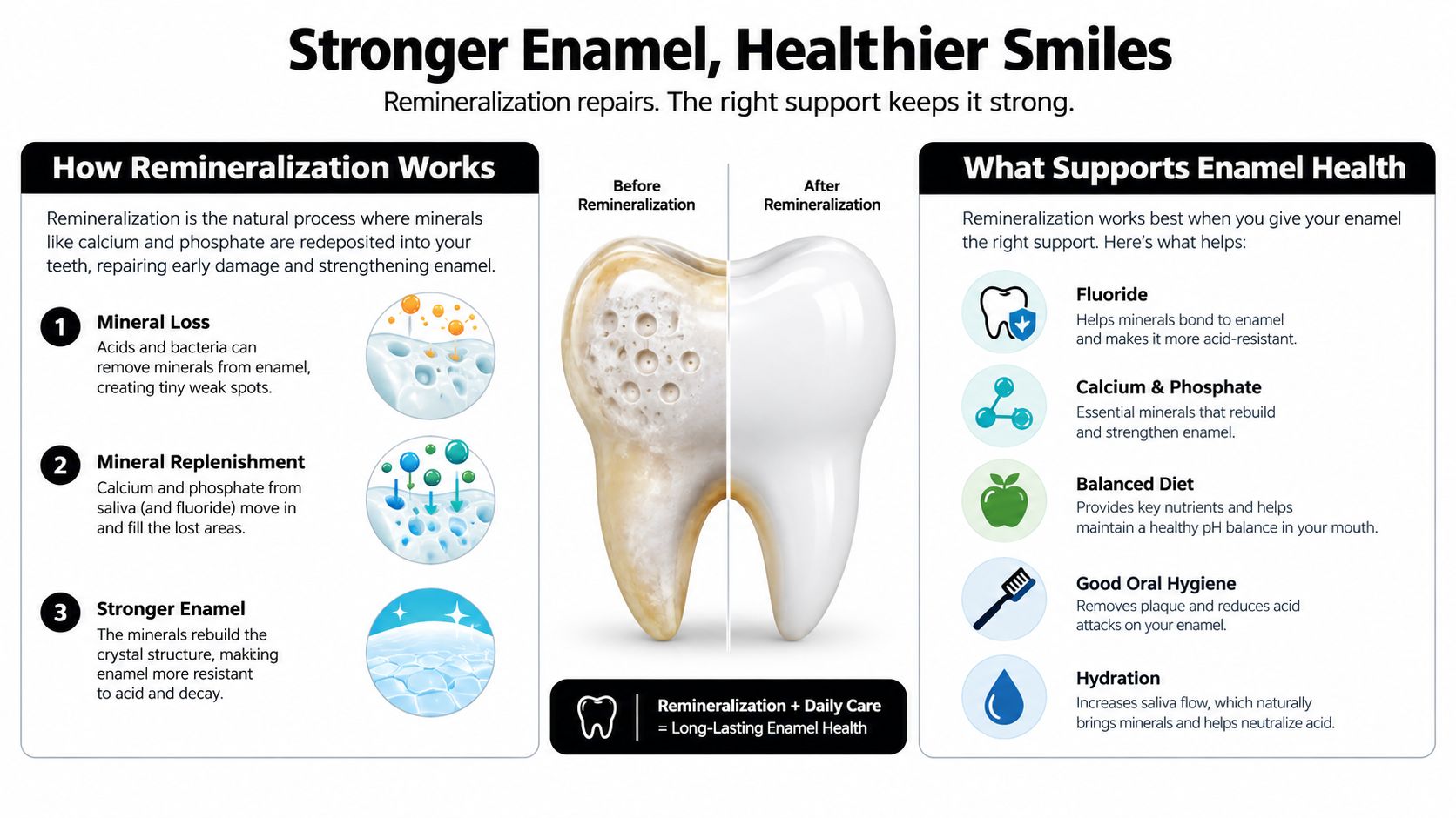
Remineralization needs three conditions working together: the mouth must move away from prolonged acidity, minerals must be available, and the enamel surface must still be capable of receiving them. Saliva supplies calcium and phosphate naturally. Topical products can add another source of mineral support or influence how enamel responds to acid.
Fluoride has the strongest evidence base for preventing, arresting, or reversing early caries lesions (the NCBI evidence summary on remineralization). It supports mineral redeposition and helps create enamel that is more resistant to acid challenges. Professional varnish is more concentrated than everyday toothpaste and should be selected with a dentist's guidance.
Nano-hydroxyapatite, often shortened to nHA, takes a mineral-based approach. Its particles are designed to resemble the calcium-phosphate mineral found in enamel, so they can support the supply of building materials at the tooth surface. The evidence is promising but mixed. An independent systematic review concluded that the available evidence was too limited and biased to establish clearly that nHA prevents caries, while newer clinical reporting includes an 18-month adult trial in which hydroxyapatite toothpaste performed similarly to 1,450 ppm fluoride toothpaste for new-decay outcomes (the systematic review of nano-hydroxyapatite toothpaste).
Remineralization support options at a glance
| Approach | How it supports remineralization | Evidence context | Best fit |
|---|---|---|---|
| Fluoride toothpaste | Supports mineral redeposition and acid resistance | Most established topical benchmark | People comfortable using fluoride |
| Professional 5% sodium fluoride varnish | Places a concentrated topical fluoride treatment on selected areas | Strong clinical support for early non-cavitated lesions | Patients with lesions identified by a dental professional |
| Nano-hydroxyapatite toothpaste | Supplies a calcium-phosphate mineral similar to enamel | Growing, mixed evidence, with important study limitations | People seeking a fluoride-free routine |
| Saliva and lower acid exposure | Helps raise pH and return minerals to enamel | Foundational biological support | Everyone, alongside brushing and dental care |
Families comparing mineral-based routines can read more about micro-hydroxyapatite toothpaste. Ingredient choice should reflect age, swallowing habits, caries risk, professional advice, and personal preferences. No toothpaste should be described as filling a cavity or regrowing missing tooth structure.
Everyday Habits That Help Prevent Decay and Support Early Repair
The most useful routine is consistent, gentle, and easy enough to repeat. Start with brushing twice daily using a toothpaste that fits your preferences and dental guidance. If you choose a fluoride-free routine, a 10% nano-hydroxyapatite formula is one mineral-based option to consider, but it shouldn't be presented as a substitute for treatment of a formed cavity.
Build the routine around plaque control
Brush every tooth surface carefully, especially along the gumline and around orthodontic brackets. Clean between teeth with floss or an interdental tool, because a toothbrush doesn't reach every contact area. A tongue scraper can help remove coating and make the mouth feel fresher, but it doesn't replace brushing or interdental cleaning.
- Use light pressure: Scrubbing harder doesn't make enamel healthier and may irritate gums.
- Clean between teeth: Choose floss picks, string floss, or interdental brushes based on what you can use reliably.
- Support children's routines: Select an age-appropriate product and supervise young children so they use it safely.
Reduce repeated acid challenges
Sugar quantity matters, but frequency also shapes how often plaque bacteria have fuel for acid production. Keep sugary foods and drinks as occasional parts of meals rather than grazing throughout the day. Acidic drinks can also challenge enamel, so water between meals is a simple upgrade.
Saliva is part of the repair environment. Staying hydrated supports normal saliva flow, while persistent dry mouth deserves discussion with a healthcare or dental professional. Avoid using abrasive whitening products casually if enamel feels sensitive or looks thinner.
For a practical routine focused on daily prevention, see this guide to how to prevent tooth decay naturally. Mouthology's 10% nano-hydroxyapatite toothpaste is one fluoride-free product option in this broader category, not a treatment for cavities.
Regular dental visits add something home care can't: trained assessment of whether a surface is intact, changing, or already cavitated. Early attention gives you more choices and less uncertainty.
Your Next Steps and When to See a Dentist
If you notice a new white spot, don't assume it's either harmless or an established cavity. Arrange a dental assessment if the mark is changing, feels rough, appears to be spreading, or comes with sensitivity. A dentist can determine whether the surface remains intact and whether monitoring, fluoride varnish, sealants, or restorative care makes sense.
Prompt care is especially important for a visible hole, persistent pain, swelling, or signs that the problem may have moved beyond the enamel. These findings don't mean you should panic. They mean the tooth needs an examination rather than a home experiment.
Ask direct questions at the appointment:
- “Is the surface intact, or is there cavitation?”
- “Could this be an early white-spot lesion or another enamel change?”
- “Would monitoring, fluoride treatment, or a sealant be appropriate?”
- “If I prefer fluoride-free care, what routine fits my risk level?”
The central answer remains reassuring and precise. Early mineral loss deserves attention because remineralization may arrest or reverse it. A cavity with surface breakdown cannot grow back and requires professional treatment.
If you've noticed a white spot, rough area, sensitivity, or possible cavity, schedule a dental examination rather than waiting for symptoms to decide for you. Use the visit to discuss the lesion's stage, your preferred fluoride or fluoride-free routine, and a practical plan for protecting the enamel you still have.
